Acute Respiratory Failure

In patients with essentially normal lung tissue, acute respiratory failure (ARF) usually means a partial pressure of arterial carbon dioxide (PaCO2) greater than 50 mm Hg and a partial pressure of arterial oxygen (PaO2) less than 50 mm Hg. These limits, however, don't apply to patients with chronic obstructive pulmonary disease (COPD), who commonly have a consistently high PaCO2 and low PaO2. In COPD patients, ARF is indicated only by acute deterioration in arterial blood gas (ABG) levels an corresponding clinical deterioration.

Causes
  • Airway irritants--smoke or fumes
  • Any condition that increases the work of breathing and decreases the respiratory drive of patients with COPD
  • Chest trauma
  • Head trauma
  • Heart failure
  • Injudicious use of sedatives, opioids, tranquilizers, or oxygen
  • Metabolic acidosis
  • Myocardial infarction
  • Myxedema
  • Pneumothorax
  • Pulmonary emboli
  • Thoracic or abdominal surgery
Signs and symptoms
  • Increased, decreased, or normal respiratory rate, depending on the cause; shallow or deep respirations, or respirations that alternate between the two; and air hunger
  • Cyanosis of the oral mucosa, lips, and nail beds possible, depending on the hemoglobin (Hb) level and arterial oxygenation
  • Crackles, rhonchi, wheezes, or diminished breath sounds
  • Restlessness, confusion, loss of concentration, irritability, tremulousness, diminished tendon reflexes, and papilledema; possible coma
  • Tachycardia, with increased cardiac output cardiac output and mildly elevated blood pressure (occurs early in response to a low PaO2)
  • Arrhythmias
  • Pulmonary hypertension
  • Accessory muscle use
  • Tachypnea
  • Cold, clammy skin
Diagnostic tests
  • Pulse oximetry may show decreased arterial oxygen saturation.
  • ABG analysis reveals hypercapnia and hypoxemia; bicarbonate levels are increased, indicating metabolic compensation for chronic respiratory acidosis.
  • Hb levels and hematocrit are abnormally low, possibly due to blood loss, indicating decreased oxygen-carrying capacity.
  • Serum electrolyte levels may indicate hypokalemia, which may result from compensatory hyperventilation--an attempt to correct alalosis; hypochloremia is common with metabolic alkalosis.
  • White blood cell count is elevated if ARF is due to bacterial infection; in certain cases of profound septicemia, the leukocyte count may be decreased.
  • Gram stain and sputum culture results can identify pathogens.
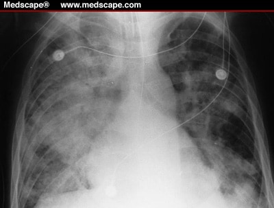
  • Chest X-rays reveal pulmonary pathology, such as emphysema, atelectasis, lesions, pneuothorax, infiltrates, or effusions.
  • Electrocardiography reveals arrhythmias, which commonly suggest cor pulmonale and myocardial hypoxia. Large P waves ("p pulmonale") may indicate a history of right-sided heart failure.
Treatment
  • Oxygen therapy using nasal prongs or a Venturi mask to raise the patient's PaO2 (if the patient has COPD) is used cautiously to promote oxygenation.
  • Bidirectional positive-pressure airway mask is used over the oronasal region or mechanical ventilation through an endotracheal or a tracheostomy tube is oredered (if significant respiratory acidosis persists).
  • High-frequency ventilation is intituted (if the patient doesn't respond to conventional mechanical ventilation.)
  • Antibiotics are prescribed for infection.
  • Bronchodilator is used to maintain airway patency.
  • Anxiolytic is ordered to promote relaxation and reduce anxiety.
  • Corticosteroids are prescribed to decrease inflammation.
  • Histamine-receptor anatgonist may be ordered to prevent ulcer formation.
Nursing considerations
  • Assess the patient's respiratory status frequently (pulse oximetry, breath sounds, and ABG results) and report changes immediately.
  • Give enough oxygen to maintain a PaO2 of at least 50 to 60 mm Hg. Patients with COPD usually require only small amounts of supplemental oxygen.
  • Maintain a patent airway. If the patient is retaining carbon dioxide, encourage him to cough and to breathe deeply with pursed lips.
  • If the patient is alert, have him use an incentive spirometer.
  • Keep the head of the bed elevated to at least 30 degrees.
  • If the patient is intubated and lethargic, turn him every 1 to 2 hours.
  • Use postural drainage and chest physiotherapy to help clear the patient's secretions.
  • Monitor and record serum electrolyte levels carefully, and correct imbalances; patient's intake and output or daily weight.
  • Monitor the patient for cardiac arrhythmias.
  • Give prescribed medications.
For mechanical ventillation
  • Check ventilator settings, cuff pressures, and ABG values frequently because the fraction of inspired oxygen (FIO2) setting depends on ABG levels. Draw a blood sample for ABG analysis 20 to 30 minutes after every FIO2 change, or check ABG levels with oximetry, as ordered.
  • Change ventilator circuits every 24 to 48 hours, per your facility's policy.
  • Suction the trachea, after hyperoxygenation, as needed. Observe for a change in the quantity, consistency, or color of secretions. Provide humidification to liquefy the secretions.
  • Keep the nasotracheal tube midline within the nostrils, and provide good hygiene. Loosen tape periodically to prevent skin breakdown. Avoid excessive movement of any tubes, and make sure the ventilator tubing is adequately supported.
  • Provide an alternate means of communication.
  • Provide emotional support to the patient and family.
read more “Acute Respiratory Failure”

Acute Respiratory Distress Syndrome

A form of pulmonary edema that causes acute respiratory failure, acute respiratory distress syndrome (ARDS) results from increased permeability of the alveolocapillary membrane. Fluid accumulates in the lung interstitium, alveolar spaces, and small airways, causing the lung to stiffen. This stiffening impairs ventilation, prohibiting adequate oxygenation of pulmonary capillary blood. Severe ARDS can cause intractable and fatal hypoxemia, but patients who recover may have little or no permanent lung damage.


Pathophysiology of ARDS

Causes
  • Aspiration of gastric contents
  • Cardiopulmonary bypass
  • Drug overdose (barbiturates, glutethimide, opioids) or blood transfusion
  • Microemboli (fat or air emboli or disseminated intravascular coagulation)
  • Near drowning
  • Oxygen toxicity
  • Pancreatitis
  • Sepsis (primarily gram-negative)
  • Smoke or chemical inhalation (nitrous oxide, chlorine, ammonia)
  • Trauma (lung contusion, head injury, long bone fracture with fat emboli)
  • Viral, bacterial, or fungal pneumonia
Signs and symptoms

Early
  • Rapid, shallow breathing and dyspnea (within hours to days of the initial injury)
  • Intercostal and suprasternal retractions
  • Crackles and rhonchi on auscultation
  • Hypoxemia causes restlessness, apprehension, mental sluggishness, motor dysfunction, and tachycardia
Late
  • Overwhelming hypoxemia
  • Hypotension
  • Decreased urine output
  • Respiratory and metabolic acidosis
  • Ventricular fibrillation or standstill
Diagnostic tests
  • Arterial blood gas (ABG) analysis first shows a decreased partial pressure of arterial oxygen (PaO2)---less than 60 mm Hg---and a decreased partial pressure of arterial carbon dioxide (PaCO2)---less than 35 mm Hg. pH ussually shows respiratory alkalosis. As ARDS becomes more severe, ABG levels indicate respiratory acidosis (a PaCO2 greater than 45 mm Hg) and metabolic acidosis (a bicarbonate level less than 22 mEq/L) aswee as decreasing PaO2, despite oxygen therapy.
  • Pulmonary artery (PA) catheterization helps identify the cause of pulmonary edema by allowing evaluation of pulmonary artery wedge pressure (PAWP) (normal PAWP values in ARDS 12 mm Hg or less) and collection of PA blood (shows decreased oxygen saturation, indicating tissue hypoxia).
  • Serial chest X-rays initially show bilateral infiltrates; in lter stages, the X-rays show ground-glass appearance and, eventually (as hypoxemia becomes irreversible), "whiteouts" of both lung fields.
  • Gram stain and sputum culture and sensitivity test results show infectious organism (if infection is the cause).
  • Blood cultures reveal the infectious organism.
  • Serum amylase levels increase if the patient has underlying pancreatitis.
  • Toxicology test identifies the drug ingested in ARDS caused by a drug overdose.
Treatment
  • Treatment of the underlying cause is started.
  • Humidified oxygen is given through a tight-fitting mask, which allows for the use of continuous positive airway pressure.
  • For hypoxemia that doesn't respond adequately to this treatment, the following may be ordered: ventilatory support with intubation, volume ventilation, and positive end-expiratory pressure (PEEP); pressure-controlled inverse ratio ventilation to reverse the conventional inspiration-to-expiration ratio and minimize the risk of barotrauma.
  • When a patient with ARDS needs mechanical ventilation, an opioid, sedative, or neuromuscular blocker may be ordered to ease ventilation and decrease oxygen consumption.
  • High-dose steroids may be prescribed for ARDS caused by fat emboli or chemical injury.
  • Diuretics are oredered.
  • Correction of electrolyte and acid-base abnormalitiesis oredered.
  • I.V. fluids and a vasopressor may be needed to maintain blood pressure.
  • Antimicrobial is prescribed if the patient has a treatable infection.
Nursing Considerations
  • Frequently assess the the patient's respiratory status. Be alert for retractions on insporation. Note rate, rhythm, and depth of respirations, and watch for dyspnea and the use of accessory muscles of respiration. On auscultation, listen for adventitious or diminished breath sounds. Check for clear, frothy sputum that may indicate pulmonary edema.
  • Observe and document the patient's neurologic status, including level of consciousness and mental sluggishness.
  • Maintain a patent airway by suctioning, using sterile, nontraumatic technique. Ensure adequate humidification to help liquefy tenacious secretions.
  • Closely monitor heart rate and blood pressure. Watch for arrhythmias that may result from hypoxemia, acid-base disturbances, or electrolyte imbalance.
  • GIve medications as prescribed.
  • With PA catheterization, know the desired PAWP level. Watch for decreasing mixed venous oxygen saturation.
  • Monitor serum electrolyte levels, and report any imbalances immediately. Measure intake and output, and weigh the patient daily.
  • Check ventilator settings frequently. Monitor ABG levels; check for metabolic and respiratory acidosis and PaO2 changes.
  • If the patient has severe hypoxemia, he may need controlled mechanical ventilation with PEEP and inverse ratio ventilation. Give sedatives, as needed, to reduce restlessness.
  • Because PEEP may decrease cardiac output, check for hypotension, tachycardia, and decreased urine output. Suction only as needed to maintain PEEP.
  • Reposition the patient frequently, and note any increase in secretions, temperature, or hypotension, which may indicate a deteriorating condition.
  • Accurately record caloric intake. Give tube feedings and parenteral nutrition, if required.
  • Perform passive range-of-motion exercises, or help the patient perform active exercises, if possible. Provide meticulous skin care. Allow periods of uninterrupted sleep.
  • Provide the patient and his family with emotional support.
read more “Acute Respiratory Distress Syndrome”

Acute Coronary Syndromes


Acute myocardial infarction (MI), including ST-segment elevation MI (STEMI) and non-ST-segment elevation MI (NSTEMI), and unstable angina are now recognized as part of a group of clinical diseases called acute coronary syndromes (ACSs). Rupture or erosion of plaque--an unstable and lipid-rich substance--initiates almost all coronary syndromes. The rupture results in platelet adhesions, fibrin clot formation, and activation of thrombin.

Mortality is high when treatment is delayed, and almost one-half of sudden deaths caused by an MI occur before hospitalization or within 1 hour of the onset of symptoms. The prognosis improves if vigorous treatment begins immediately.

Causes
  • Atherosclerosis
  • Embolus
Risk factors
  • Diabetes
  • Elevated homocysteine, C-reactive protein, and fibrin levels
  • Excessive alcohol consumption
  • Family history of heart disease
  • High-fat, high-carohydrate diet
  • Hyperlipoproteinemia
  • Hypertension
  • Obesity
  • Postmenopausal status
  • Sedentary lifestyle
  • Smoking
  • Stress
Signs and symptoms

Angina
  • Burning, squeezing, and crushing tightness in the substernal or precordial chest that may radiate to the left arm or shoulder blade, the neck, or the jaw
  • Pain after physical exertion, emotional excitement, exposure to the cold or consumption of a large meal
MI
  • Uncomfortable pressure, squeezing, burning, severe persistent pain or fullness in the center of the chest lasting several minutes (usually longer than 15 minutes)
  • Pain radiating to the shoulders, neck, arms, or jaw or pain in the back between the shoulder blades
  • Lightheadedness or fainting
  • Sweating
  • Nausea
  • Shortness of breath
  • Anxiety or a feeling of impending doom
Diagnostic tests
  • Electrocardiography helps determine which area of the heart and which coronary arteries are involved.
  • Serial cardiac enzymes and protein levels may show a characteristic rise in CK-MB, the proteins troponin T and I, and myoglobin.
  • Laboratory testing may reveal elevated white blood cell count and erythrocyte sedimentation rate and changes in electrolyte levels.
  • Echocardiography may show ventricular wall motion abnormalities and may detect septal or papillary muscle rupture.
  • Transesophageal echocardiography may reveal areas of decreased heart muscle wall movement, indicating ischemia.
  • Chest X-rays may show left-sided heart failure, cardiomegaly, or other noncardiac causes of dyspnea and chest pain.
  • Nuclear imaging scanning using thallium 201 or technetium 99m can be used to identify areas of infarction and areas of viable muscle cells.
  • Cardiac catheterization may be used to identify the involved coronary artery as well as to provide information on ventricular function and pressures and volumes within the heart
Viewing the coronary arteries

Acute coronary syndrome commonly results when a thrombus progresses and occludes blood flow through a coronary artery. This illustration shows the major coronary vessels that may be involved.

  • For the patient with STEMI, treatment includes the above initial interventions and also:
  1. thrombolytic therapy (unless contraindicated) within 12 hours of onset of symptoms to restore vessel patency and minimize necrosis.
  2. heparin I.V. to promote patency in the affected coronary artery.
  3. a glycoprotein IIb/IIa inhibitor to minimize platelet aggregation.
  4. an angiotensin-converting enzyme (ACE) inhibitor to reduce afterload and preload and prevent remodeling (begin 6 hours after admission or when the patient's condition is stable)
  5. PTCA, stent placement, or CABG surgery to open blocked or narrowed arteries.
Nursing considerations
  • Institute continuous cardiac monitoring and frequently monitor the electrocardiogram (ECG) to detect rate changes or arrhythmias. Place rhythm strips in the patient's chart periodically according to yourfacility's policy.
  • Monitor and record the patient's blood pressure, temperature, and heart and breath sounds.
  • Explain the importance of reporting pain immediately.
  • Obtain a 12-lead ECG during episodes of chest pain.
  • Assess and record location, severity, and duration of pain.
  • Give prescribed analgesics and other medications.
  • Check the patient's blood pressure after giving nitroglycerin, especially after the first dose.
  • Monitor intake and output closely.
  • Monitor the patient for crackles, cough, tachypnea, and edema, which may indicate impending left-sided heart failure.
  • If the pateint has undergone PTCA, provide sheath care. Maintain strict bed rest and keep the leg with the sheath insertion site immobile. Monitor the site closely for bleeding. Check peripheral pulses in the affected leg frequently.
  • Provide emotional support, and help to reduce stress and anxiety.
  • Initiate cardiac rehabilitation, according to your facility's protocol.
  • Review dietary restrictions with the patient. If he must follow a low-cholesterol, low-sodium, low-fat, high-fiber diet, provide a list of foods that he should avoid. Ask the dietitian to speak to the patient and his family.
  • Refer the patient to a smoking-cessation program, if needed.
  • Thoroughly explain the patient's treatment regimen. Warn the patient about adverse reactions to drugs, and advise him to report them to his practitioner.
  • Refer the patient to a weight-reduction program, if needed.
  • Counsel the patient to resume sexual activity progressively.
read more “Acute Coronary Syndromes”

Acromegaly and Gigantism


Acromegaly (above picture) and gigantism (below picture) are chronic, progressive diseases marked by hormonal dysfunction and startling skeletal overgrowth. Acromegaly occurs after epiphyseal closure, causing bone thickening and transverse growth and visceromegaly. Gigantism begins before epiphyseal closure and causes proportional overgrowth of all body tissues. Acromegaly develops slowly, whereas gigantism develops abruptly. Although the prognosis depends on the on the causative factor, these disorders usually reduce life expectancy unless treated in a timely way.


Causes
  • Extrapyramidal pituitary lesions or other tumors that cause oversecretion of hGH
  • Oversecretion of human growth hormone (hGH) that produces changes throughout the entire body, resulting in acromegaly and, when oversecretion occurs before puberty, gigantism
  • Possible genetic cause
  • Somatotropic adennomas
Signs and Symptoms

Acromegaly
  • Arthropathy
  • Carpal tunnel syndrome
  • Proximal muscle weakness
  • Fatigue
  • Acanthosis nigricans (an eruption of velvet benign growths and hyperpigmentation occurring in the skin of the axillae, neck, anogenital area)
  • Skin tags
  • Oily skin
  • Cartilaginous and connective tissue overgrowth
  • Enlarged supraorbital ridge and thickened ears and nose
  • Marked projection of the jaw, which may interfere with chewing
  • Voice sounds deep and hollow
  • Thickened fingers
  • Coronary artery disease
  • Cardiomyopathy with arrhythmias, left ventricular hypertrophy, and decreased diastolic function
  • Hypertension
  • Upper airway obstruction with sleep apnea
  • Generalized visceromegaly, including cardiomegaly, macroglosia, and thyroid gland enlargement
  • Barrel chest and kyphosis
  • Signs of diabetes mellitus and glucose intolerance
Gigantism
  • Same skeletal abnormalities and signs of glucose intolerance seen in acromegaly
  • Pituitary tumor enlargement (causing loss of other trophic hormones, such as thyroid-stimulating hormone, lutenizing hormone, follicle-stimulating hormone, and corticotropin)
Diagnostic tests
  • Serum hGH levels measured by radio-immunoassay typically are elevated.
  • Glucose suppression test fails to suppress the hormone level to below the accepted normal value of 2 ng/ml.
  • Skull X-rays, a computed tomography scan, arteriography, and magnetic resonance imaging determine the presence and extent of the pituitary lesion.
  • Bone X-rays show a thickening of the cranium (especially of frontal, occipital, and parietal bones) and of the long bones as well as osteoarthritis in the spine.
Treatment
  • Cranial or transphenoidal hypophysectomy or pituitary radiation therapy is oredered to remove the underlying tumor.
  • Replacement of thyroid and gonadal hormones and cortisone are ordered, postoperatively.
  • Bromocriptine (Parlodel) and octreotide (Sandostatin) are used to inhibit hGH synthesis.
  • Provide the patient with emotional support to help him cope with an altered body image.
  • Assess the patient for skeletal changes and muscle weakness.
  • Perform or assist with range-of-motion exercises.
Do's & Dont's

Keep the patient's skin dry. Avoid using an oily lotion because the skin is already oily.

  • Monitor the patient's blood glucose level. Check for signs and symptoms of hyperglycemia, including fatigue, polyuria, and polydispsia.
  • Reassure the patient and his family that the disease causes mood changes, which can be managed with treatment.
  • Before surgery, reinforce what the surgeon has told the patient and try to allay the patient's fear.
  • After surgery
  1. Diligently monitor the patient's vital signs and neurologic status. Be alert for signs of increased intracranial pressure.
  2. Check blood glucose levels frequently. Remember, hGH levels usually fall rapidly after surgery, removing an insulin anatagonist effect in many patients and possibly precipitating hypoglycemia.
  3. Measure intake and output hourly and watch for large increases in urine output. Transient diabetes insipidus, which sometimes occurs after surgery for hyperpituitarism, can cause such increases in urine output.
  4. If the transphenoidal approach is used, the surgical site is packed with a piece of tissue usually taken from a midthigh donor site. Watch for cerebrospinal fluid leaks from the packed site. Look for increased external nasal drainage into the nasopharynx.
  5. Help the patient get out of bed and walk on the first or second day after surgery, per prescribed level of activity.
  • Make sure the patient and his family understand which hormones are to be taken and why, as well as the correct times and dosages.
read more “Acromegaly and Gigantism”

Acne Vulgaris

An inflammatory disorder of the sebaceous glands, acne vulgaris is the most common skin problem in adolescents, although lesions can appear as early as age 8. Although acne is more common and more severe in boys than girls, it usually occurs in girls at an earlier age and tends to last longer, sometimes into adulthood. The prognosis is good with treatment.


Causes
  • Exact cause unknown
  • Possible primary causes: follicular occlusion, androgen-stimulated sebum production, and Propionibacterium acnes
Flare-ups
  • Certain drugs, including corticosteroids, glucocorticoids, halogens, phenobarbital, phenytoin (Dilantin), isoniazid (Laniazid), and lithium
  • Cosmetics
  • Emotional stress
  • Exposure to industrial compounds
  • Trauma or rubbing from tight clothing
  • Unfavorable climate
Signs and symptoms
  • Closed comedo, or whitehead (if it doesn't protrude from the follicle and is covered by the epidermis)
  • Open comedo, or blackhead (if it does protrude and isn't covered by the epidermis)
  • Inflammation and characteristic acne pustules, papules or, in severe forms, cysts or abscesses
  • Scarring, if chronic recurring lesions
Diagnostic tests
  • Results of culture and sensitivity testing of pustules show causative organism of secondary bacterial infection.
Treatment
  • A topical antibacterial (such as benzoyl peroxide, clindamycin [Cleocin], or erythromycin) is prescribed, alone or with tretonin (Avita), a keratolytic, or salicylic acid.
  • A systematic antibiotic, usually tetracycline (Sumycin), decrease bacterial growth until the patient is in remission; then a lower dose is used for long-term maintenance. Tetracyline is contraindicated during pregnancy and childhood because it discolors developing teeth. Erythromycin is an alternative for these patients.
  • Because of its severe adverse effects, a 16 to 20-week course of oral isotretinoin (Accutane) is limited to those with severe papulopastular or cystic acne who don't respond to conventional therapy.
Avoiding risks of isotretinoin therapy

Because isotretinoin is known to cause birth deftects, the manufacturer, with approval of the Food and Drug Administration, recommends th following precautions:
  • Pregnancy testing before dispensing
  • Effective contraception during treatment
  • Dispensing only a 30-day supply
  • Repeat pregnancy testing throughout the treatment period
  • Informed consent of the patient or parents regarding the drug's adverse effects

  • Oral hormonal contraceptives (such as Ortho Tri-Cyclen) or spironolactone (Aldactone) may be prescribed for female patients because these drugs have anti-androgenic effects.
  • Intralesional cortecosteroid injections may be oredered.
  • Exposur to ultraviolet light may be ordered (but never when a photosensitizing agent, such a tretinoin, is being used).
  • Cyrotherapy may be ordered.
  • Additional treatments include comedo extraction, which involves drainage and extraction of larger cysts.
Nursing Considerations
  • Check the patient's drug history because certain drugs may cause an acne flare-up.
  • Try to identify predisposing factors that may be eliminated or modified.
  • Explain to the patient and his family that the prescribed treatment is more likely to improve acne than a strict diet and fanatic scrubbing with a soap and water. Provide written instructions regaring treatment.
  • Intsruct the patient using tretinoin to apply it at least 30 minutes after washing the face and at least 1 hour before bedtime. Warn against using it around the eyes or lips. After treatments, the skin should look pink and dry. If it appears red or starts to peel, the preparation may have to be weakened or applied less often. Advise the patient to avoid exposure to sunlight or ro use a sunscreen.
  • If the prescribed regimen includes tretinoin and benzoyl peroxide, avoid skin irritation by using one preparation in the morning and the other at night.
Drug Challenge

Instruct the patient to take tetracycline on an empty stomach and not to take it with antacid or milk.

  • If the patient is taking isotretinoin, tell him to avoid vitamin. A supplements, tell him to avoid vitamin A supplements, which can worsen any adverse reactions.
  • Warn the female patient taking isotretinoin about the severe risk of terato genesis associated with the use of this drug.
  • Monitor the patient's liver function and lipid levels when isotretinoin is used.
  • Offer emotional support.

read more “Acne Vulgaris”